Pig Imaging Group
Welcome to the
PIG IMAGING GROUP
Our research group utilizes the pig to investigate how environmental and biological factors influence neurodevelopment.
Come check out our many neuroimaging resources and discoveries!
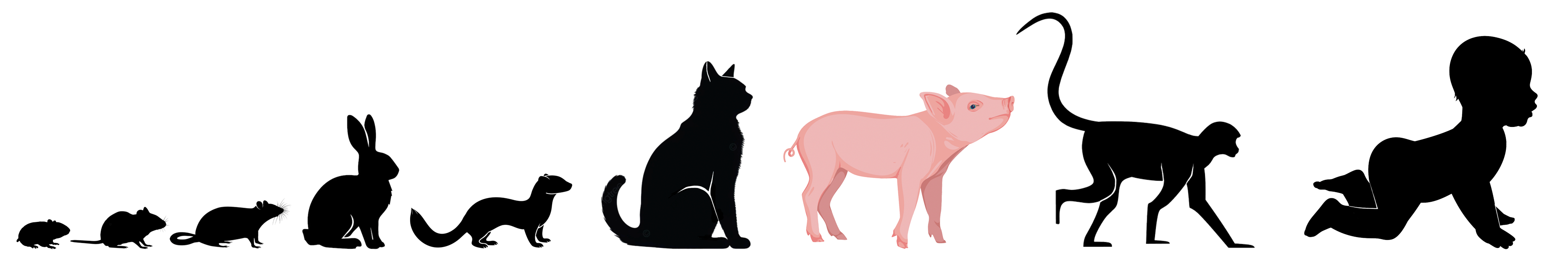
WHY PIGS?
For decades, pigs have been utilized as an animal model in neuroscience, nutrition, and development, but why is that? How close is a pig to a human?
Check out our work to answer those questions and
discover our neurodevelopmental translational timeline between species!
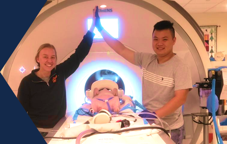
NEUROIMAGING RESOURCES
With over 1,000 individual MRI scans of the pig brain performed since 2010, our lab is well versed in multimodal imaging across multiple ages and field strengths (3T and 7T).
Utilizing these scans and with the help of Dr. Brad Sutton
from the Biomedical Imaging Center at the Beckman Institute, we have developed
multiple free open-access MRI resources for the pig.
PIG BRAIN ATLAS
Download atlases specifically made for the domesticated pig to isolate brain regions, segment the brain, and more!
pigBET SOFTWARE
An automated skull-stripping software to make neuroimaging processing a breeze. Access and download the repository for your specific uses.
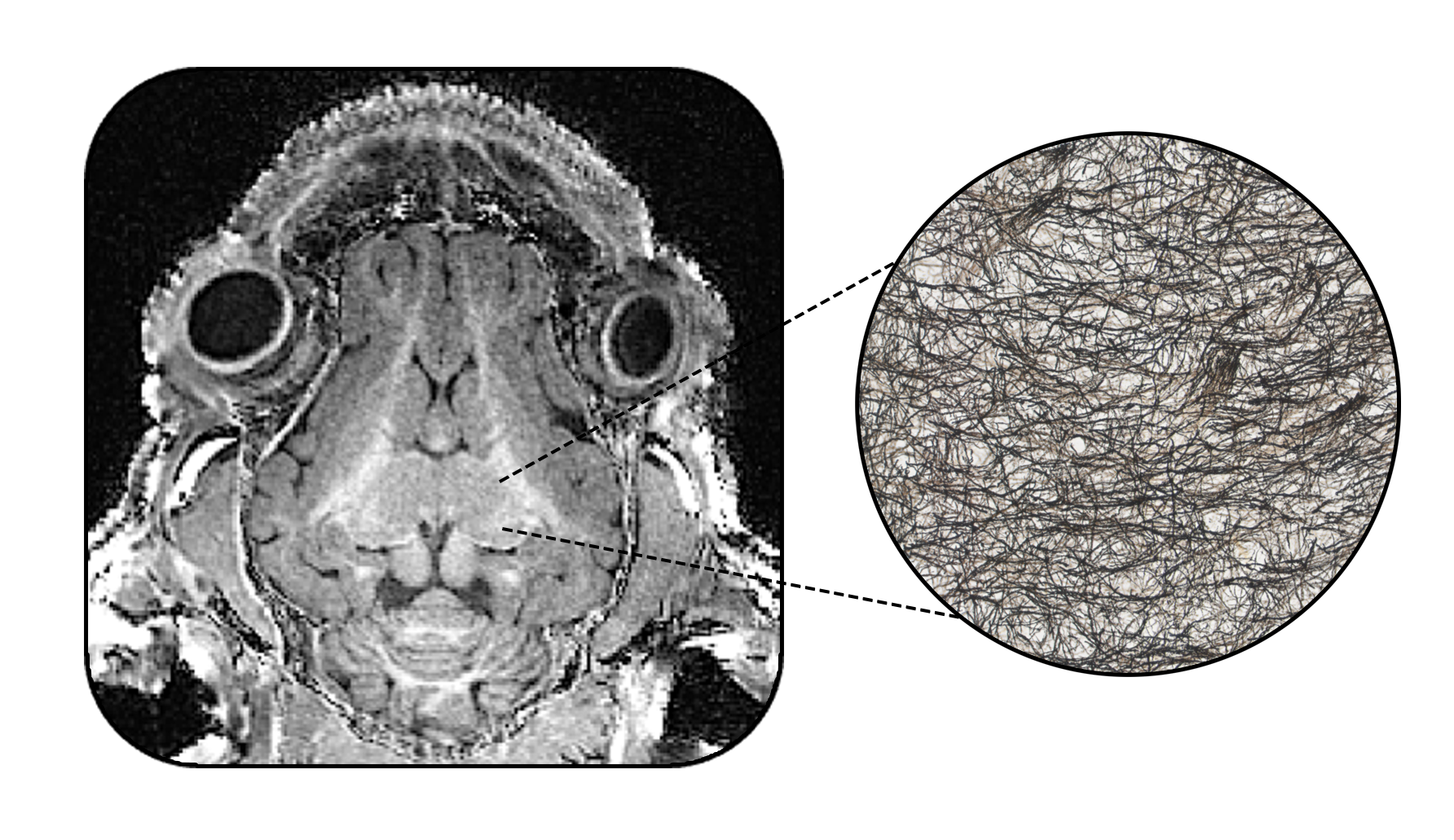
A DEEPER DIVE
What are MRI images telling us? Are scans truly reflective of biology?
To answer this, our lab paired multimodal neuroimaging together with histology to get
a deeper look into the brain.
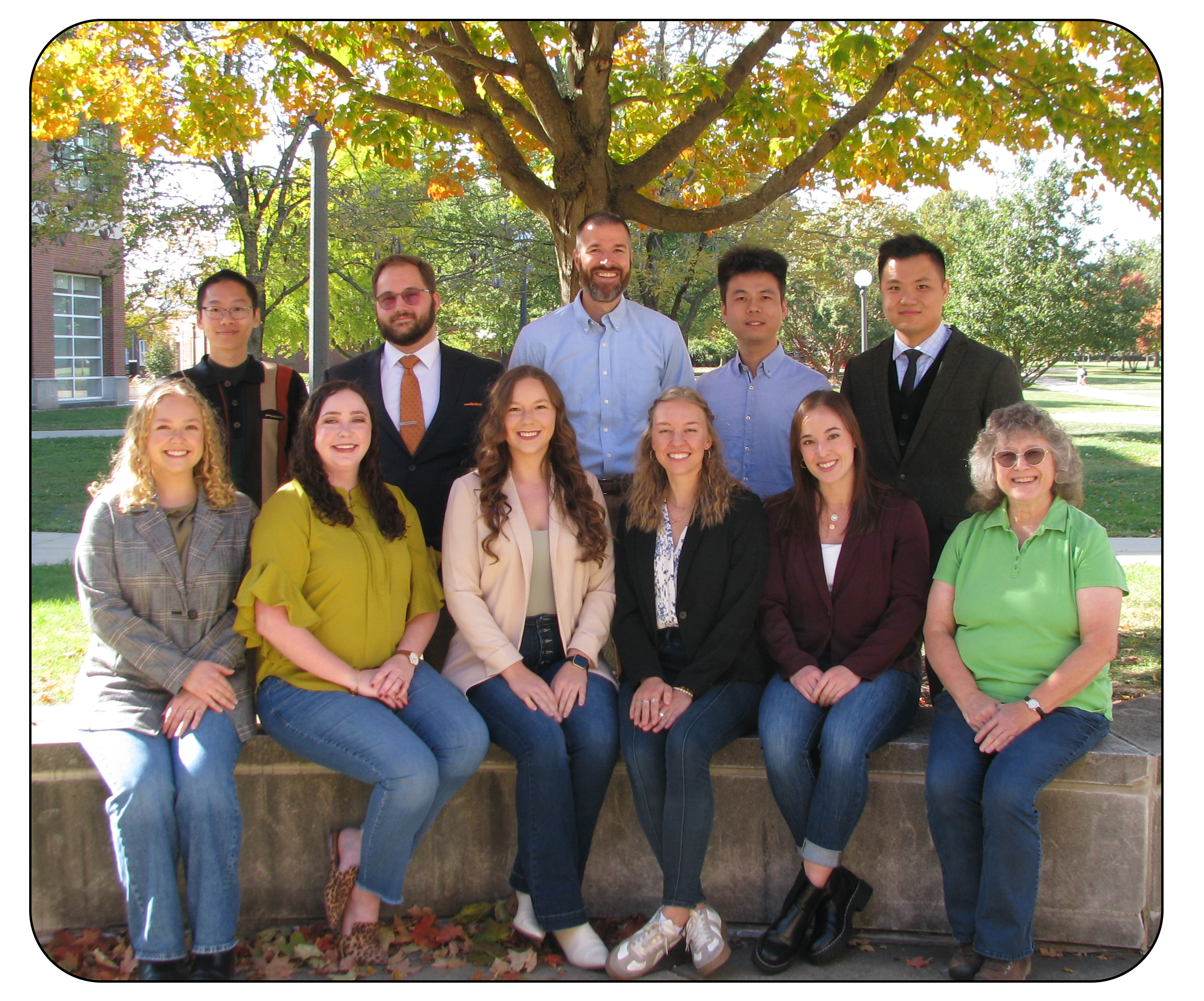
Meet our Collaborative Team
These projects would not have been possible without the collaboration between the Beckman institute for Advanced Science and Technology and ACES Department of Animal Sciences at the University of Illinois Urbana-Champaign.
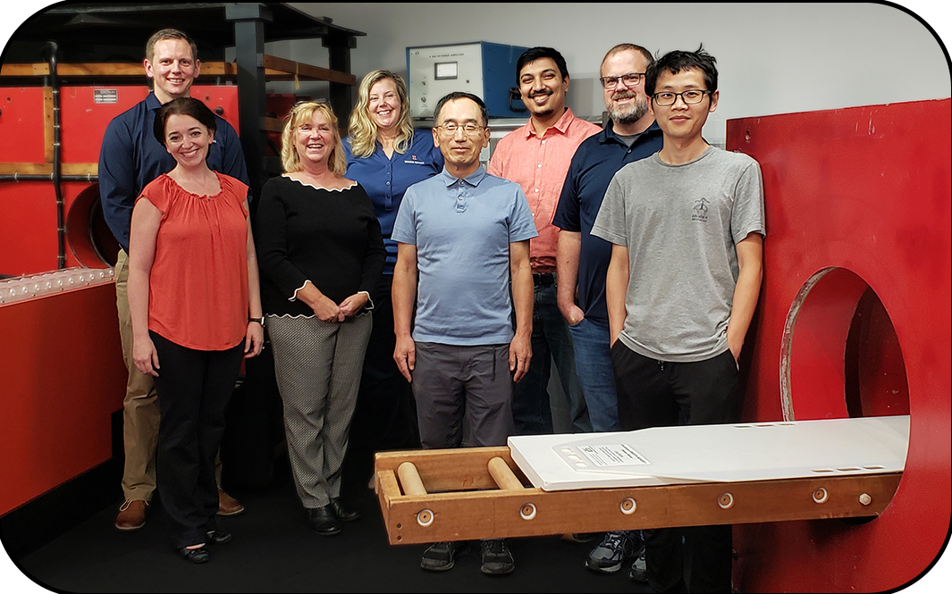
The Beckman Institute Biomedical Imaging Center. Back row: Aaron Anderson, Holly Keleher, Shreyan Majumdar and Brad Sutton. Front row: Hillary Schwarb, Tracey Wszalek, Kwanjin Jung, and Yuhui Chai.
The Beckman Institute Biomedical Imaging Center (BIC)
The Dilger Lab
Pig Imaging Group